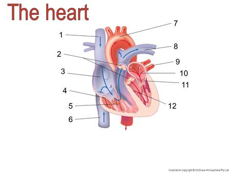
38 label the parts of a heart
Heart Labeling Flashcards | Quizlet Parts of the heart Terms in this set (11) Superior Vena Cava Inferior Vena Cava Right Atrium Tricuspid Valve Right Ventricle Pulmonary Artery Pulmonary Veins Left Atrium Bicuspid Valve Left Ventricle Aorta Students also viewed heart labeling 14 terms Images kristiquaglieri Label the Heart 15 terms Diagram bluesas9 heart labeling 19 terms Diagram AI song featuring fake Drake and Weeknd vocals pulled from streaming ... A song featuring AI-generated vocals purporting to be Drake and the Weeknd has been pulled from streaming services by Universal Music Group (UMG) after going viral over the weekend. The label ...
Heart: Anatomy and Function - Cleveland Clinic The parts of your heart are like the parts of a house. Your heart has: Walls. Chambers (rooms). Valves (doors). Blood vessels (plumbing). Electrical conduction system (electricity). Heart walls Your heart walls are the muscles that contract (squeeze) and relax to send blood throughout your body.
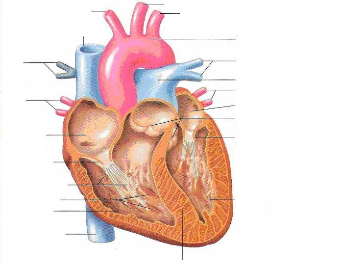
Label the parts of a heart
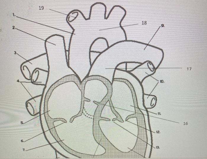
Diagrams, quizzes and worksheets of the heart | Kenhub Worksheet showing unlabelled heart diagrams. Using our unlabeled heart diagrams, you can challenge yourself to identify the individual parts of the heart as indicated by the arrows and fill-in-the-blank spaces. This exercise will help you to identify your weak spots, so you'll know which heart structures you need to spend more time studying ... The Anatomy of the Heart, Its Structures, and Functions - ThoughtCo The heart wall consists of three layers: Epicardium: The outer layer of the wall of the heart. Myocardium: The muscular middle layer of the wall of the heart. Endocardium: The inner layer of the heart. Cardiac Conduction Cardiac conduction is the rate at which the heart conducts electrical impulses. A Labeled Diagram of the Human Heart You Really Need to See The human heart, comprises four chambers: right atrium, left atrium, right ventricle and left ventricle. The two upper chambers are called the left and the right atria, and the two lower chambers are known as the left and the right ventricles. The two atria and ventricles are separated from each other by a muscle wall called 'septum'.
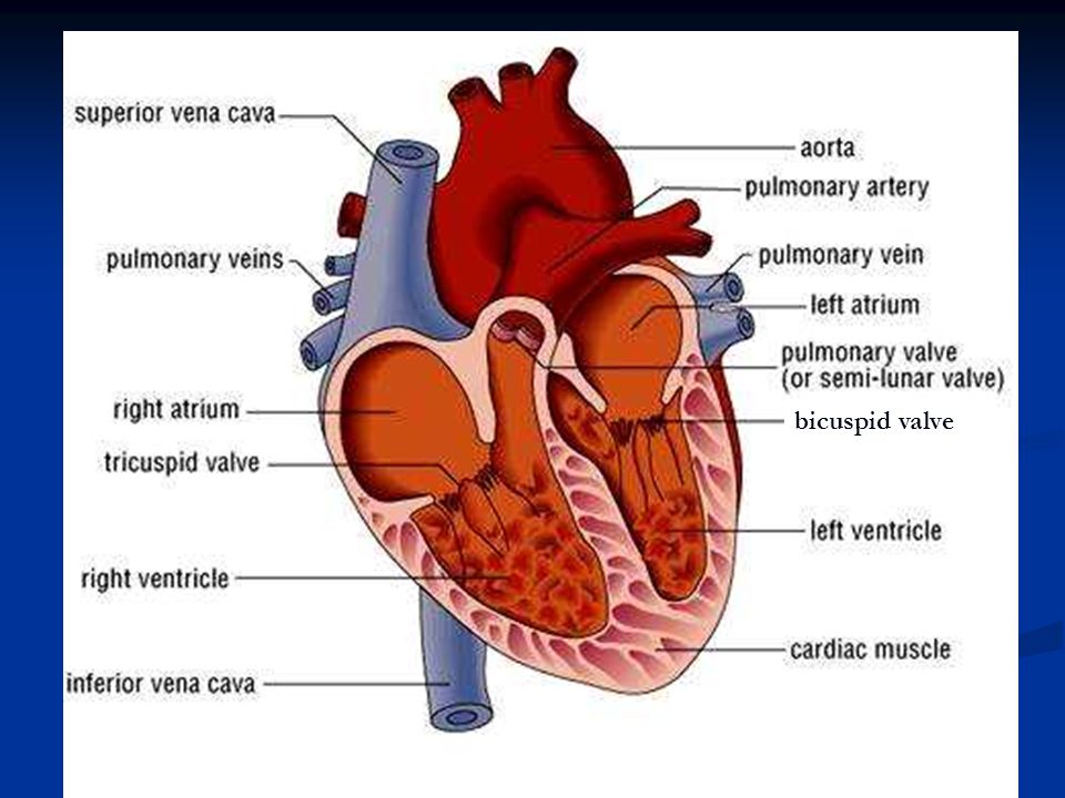
Label the parts of a heart. Human Anatomy: Label the Heart Worksheets in 3 Differentiated Levels These Human Anatomy: Label the Heart Worksheets are an excellent activity for your students to complete as part of their study of the human body.. Check out these Human Anatomy Heart resources . Exploring the Circulatory System with About Kid's Health. How to Feel Your Heart Beat with SciShowKids. If your kids learn best with songs, listen to this Song from KLT (the song is catchy, but the ... Heart anatomy: Structure, valves, coronary vessels | Kenhub Heart anatomy. The heart has five surfaces: base (posterior), diaphragmatic (inferior), sternocostal (anterior), and left and right pulmonary surfaces. It also has several margins: right, left, superior, and inferior: The right margin is the small section of the right atrium that extends between the superior and inferior vena cava . Picture of the Heart - WebMD The heart is a muscular organ about the size of a fist, located just behind and slightly left of the breastbone. The heart pumps blood through the network of arteries and veins called the... Label parts of the heart - Liveworksheets Label parts of the heartDrag and drop the labels to the correct parts indicated on the heart diagram. ID: 832107. Language: English. School subject: Biology. Grade/level: GCSE. Age: 12-18. Main content: Label parts of the heart. Other contents: Add to my workbooks (517)
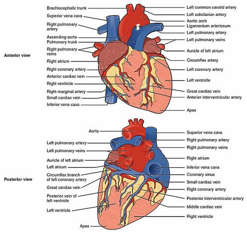
Label the heart — Science Learning Hub Label the heart Interactive Add to collection In this interactive, you can label parts of the human heart. Drag and drop the text labels onto the boxes next to the diagram. Selecting or hovering over a box will highlight each area in the diagram. aorta left ventricle pulmonary vein right atrium semilunar valve left atrium right ventricle vena cava Labeling the parts of the heart. Flashcards | Quizlet Right Atrium Identify g Left Atrium Identify t Pulmonary Trunk Identify e Left Pulmonary Artery Identify r Right Pulmonary Artery Identify c Right Ventricle Identify j Left Ventricle Identify x Inferior Vena Cava Identify m Superior Vena Cava Identify b Acending Aorta Identify d Aortic Arch Identify p Apex Identify aa Left Pulmonary Veins Labelling the heart — Science Learning Hub identify the main parts of a heart describe the functions of the different parts of the heart. This online interactive supports students to identify the different parts of the heart and the role they have in blood circulation. It can be used as both a formative and summative tool for learning. Topics Concepts Citizen science Teacher PLD Glossary Heart | Structure, Function, Diagram, Anatomy, & Facts The heart consists of several layers of a tough muscular wall, the myocardium. A thin layer of tissue, the pericardium, covers the outside, and another layer, the endocardium, lines the inside. The heart cavity is divided down the middle into a right and a left heart, which in turn are subdivided into two chambers.
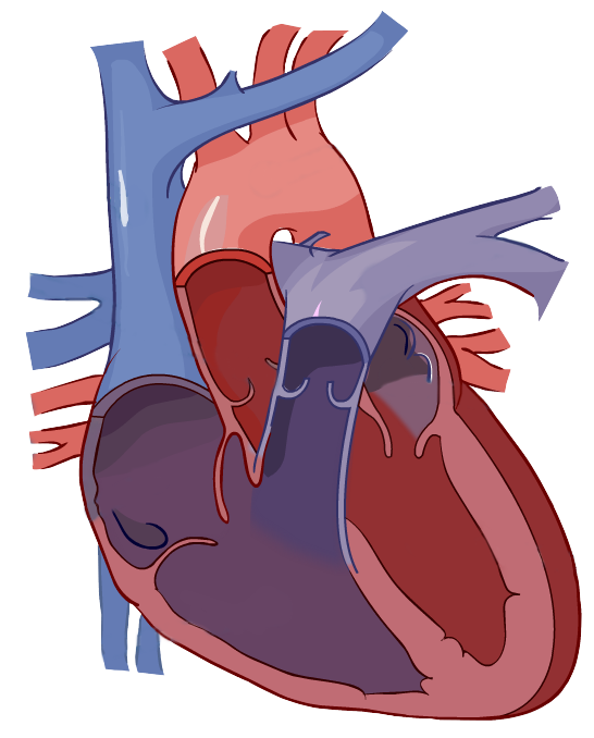
How to Draw a Human Heart: An Easy Step-By-Step Guide - WikiHow The heart works like a pump and beats 100,000 times a day. The heart has two sides, separated by an inner wall called the septum. The right side of the heart pumps blood to the lungs to pick up oxygen. The left side of the heart receives the oxygen-rich blood from the lungs and pumps it to the body. Thanks! We're glad this was helpful. A Labeled Diagram of the Human Heart You Really Need to See The human heart, comprises four chambers: right atrium, left atrium, right ventricle and left ventricle. The two upper chambers are called the left and the right atria, and the two lower chambers are known as the left and the right ventricles. The two atria and ventricles are separated from each other by a muscle wall called 'septum'. The Anatomy of the Heart, Its Structures, and Functions - ThoughtCo The heart wall consists of three layers: Epicardium: The outer layer of the wall of the heart. Myocardium: The muscular middle layer of the wall of the heart. Endocardium: The inner layer of the heart. Cardiac Conduction Cardiac conduction is the rate at which the heart conducts electrical impulses. Diagrams, quizzes and worksheets of the heart | Kenhub Worksheet showing unlabelled heart diagrams. Using our unlabeled heart diagrams, you can challenge yourself to identify the individual parts of the heart as indicated by the arrows and fill-in-the-blank spaces. This exercise will help you to identify your weak spots, so you'll know which heart structures you need to spend more time studying ...
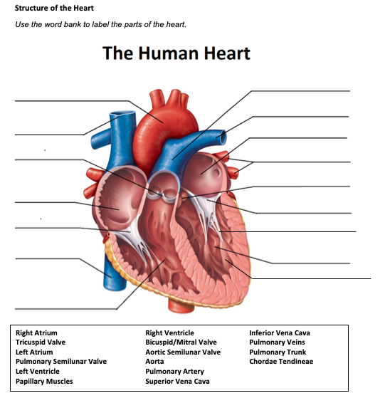
Solved Structure of the Heart Use the word bank to label the ...

Heart Anatomy: Labeled Diagram, Structures, Blood Flow ...
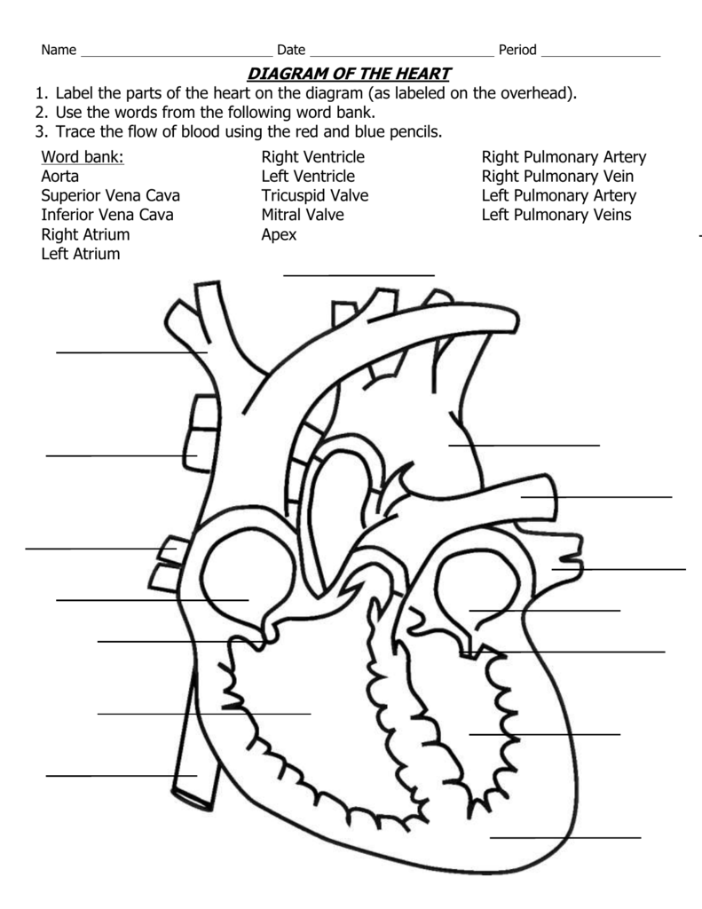
DIAGRAM OF THE HEART
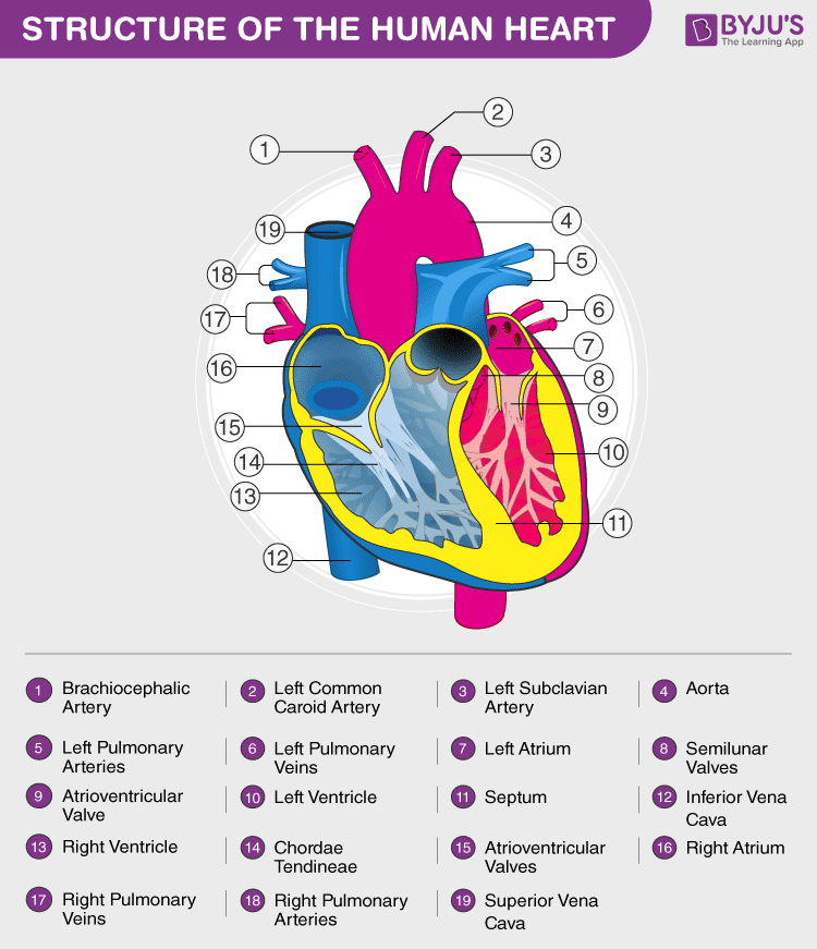
Heart Diagram with Labels and Detailed Explanation

Label the parts of the human heart

SOLUTION: Human anatomy the heart labelling practice and ...

Label the parts of the human heart and its function

Heart Anatomy Vector Illustration Labeled Organ Structure ...

The Heart and Circulatory System - ppt download
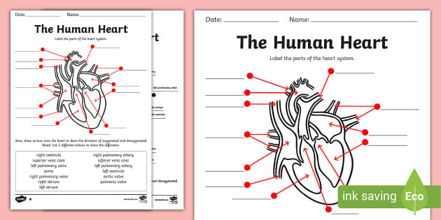
Human Heart Diagram Without Labels - Labelling Worksheet
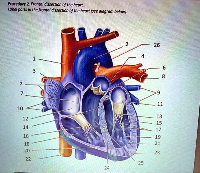
SOLVED: Procedure 2 Frontal dissection of the heart: Label ...

DIAGRAM OF THE HEART 1. Label the parts of the heart on the ...

File:Diagram of the human heart.svg - Wikimedia Commons
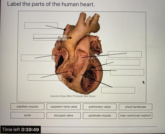
Solved Label the parts of the human heart. Dicson Shawn ...
Label the heart — Science Learning Hub

pictures with parts labeled - Google Search | Human heart ...

16,090 Human Heart Diagram Images, Stock Photos & Vectors ...
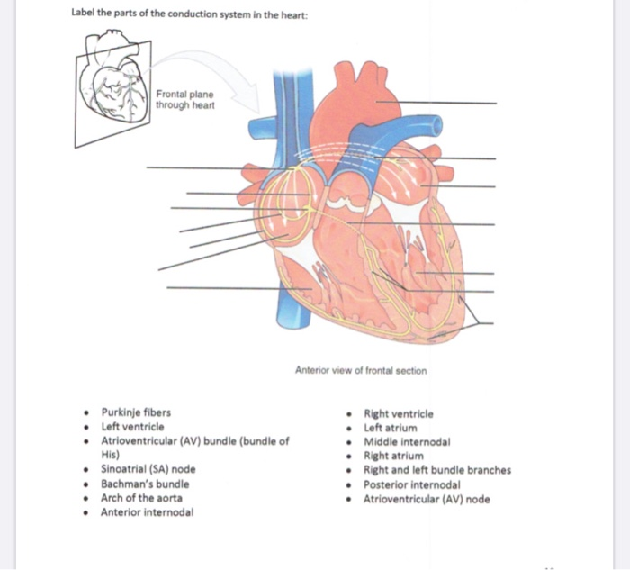
Solved Label the parts of the conduction system in the ...

Draw a diagram of the sectional view of human seminiferous ...

Sketch the internal structure of human heart. Label all the ...

Label the parts of human heart.

Heart Diagram | Heart diagram, Parts of the heart, Printable ...
The Structure of the Heart Learning Objectives: Label the ...

Q1 Given alongside is a diagram of human heart showing its ...
Anatomy of the Human Heart - Physiopedia
Solved Label the parts of the heart that correspond to the ...

Q4 Given alongside is a diagram of the human heart showing ...

label the parts of a heart - Brainly.ph

Heart parts interactive worksheet

Human heart diagram hi-res stock photography and images - Alamy
Label the heart — Science Learning Hub
Label the Heart Quiz

File:Heart diagram-en.svg - Wikipedia
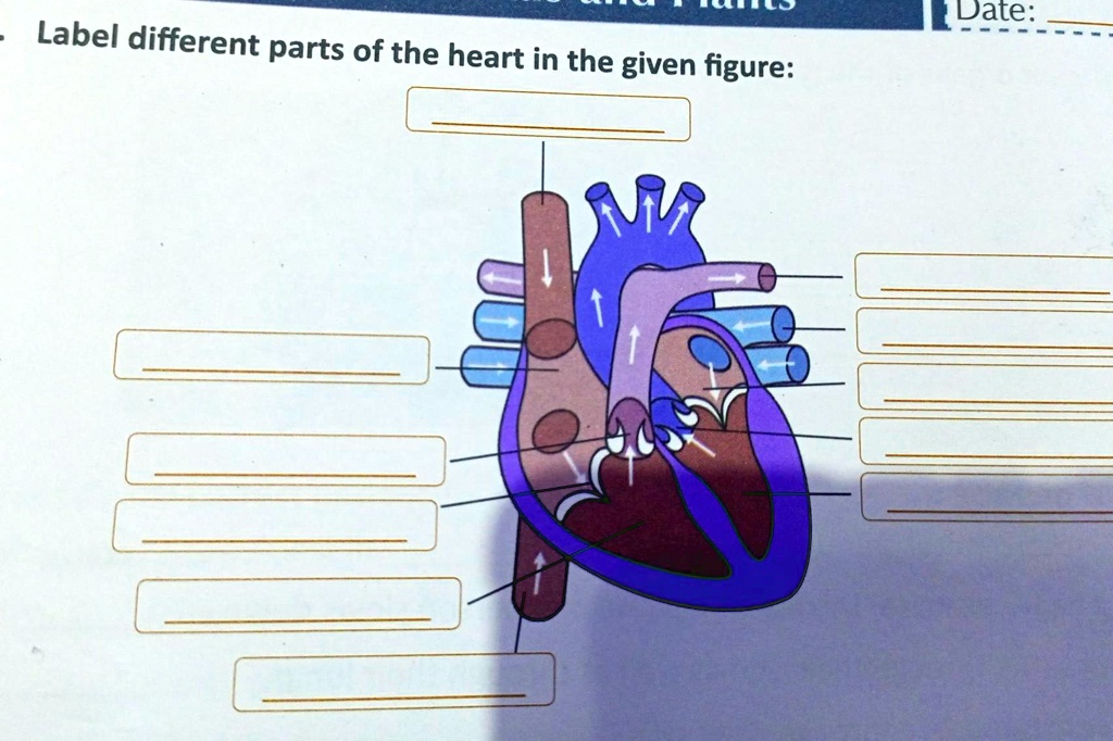
SOLVED: "1. Label different parts of the heart in the given ...
The Structure of the Heart Learning Objectives: Label the ...

The Parts of the Heart (Blank) Printable Printable (6th ...

Label parts of the heart worksheet
Solved] Correctly label the following parts of the heart ...
Post a Comment for "38 label the parts of a heart"